Aurox used specially designed sCMOS cameras from XIMEA to develop an all-in-one microscope
Aurox's unity – New development in laser free confocal microscopes
Confocal microscopy is a well known and popular approach improving the conventional widefield optical microscopy with several major advantages.
Modern confocal microscopes are fully integrated digital systems consisting of detectors, computer, laser and a beam scanning assembly.
It is a relatively sophisticated task to come up with novel functionality that would benefit the customers using these systems in various scientific fields.
Aurox is an optical imaging manufacturer based in UK with an experienced team of experts successfully offering such advancements for over 15 years.
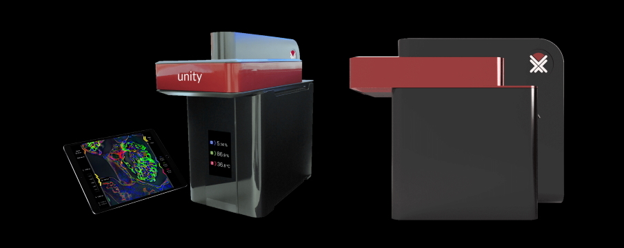
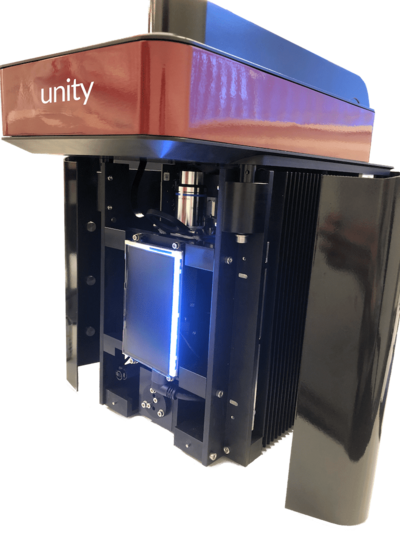
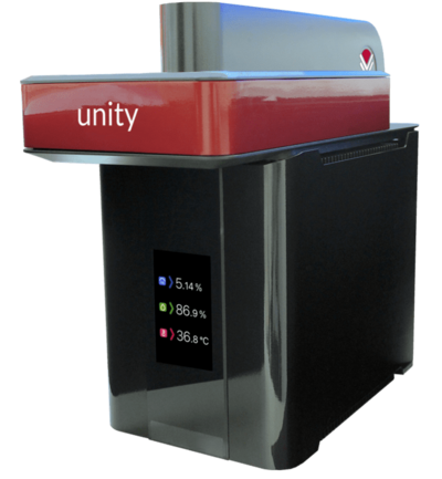
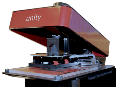
Their latest system called Unity makes confocal microscopy faster, easier, more affordable and accessible for wet lab biologists and medical researchers.
To finalize it for mass market the newest technologies were required allowing to miniaturize the components thus making the end product extremely compact and self contained.
For this purpose, the Aurox team decided to collaborate with an innovator who was able to provide an OEM solution suitable for the demanding goals.
When a unique approach and agile implementation are important, XIMEA is eager to help with the experience and the latest technological achievements.
Introducing Aurox
A leader in the design and manufacture of innovative optical imaging equipment, Aurox has received multiple business and technology awards including, the Queen's Award for Enterprise, the Institute of Physics (IOP) Innovation award and the R&D100 award.
Aurox's leading product is the Clarity Laser Free Confocal (LFC) unit, which is based on the award winning Aurox SD62 and delivers affordable confocal microscopy to a wider user community through its ability to attach to any conventional microscope and to provide high quality 3D images acquired in real time.
Aurox further enhanced the success of the Clarity with Unity, decreasing the size, adding new functionality and feature set, making the process faster and simple.
Previous systems as well as Unity address specific market needs in applications ranging from *live cell imaging* and high throughput screening to materials inspection.
Picture 1: Unity - a compact bench top microscope
Measurement principle | Confocal fluorescence microscope - laser free |
Confocality | 0.6 μm Full width at half maximum (FWHM) with 1.4 N.A oil immersion objective |
Disc | 1 sector structured illumination disc |
Sample type | Microscope slides, Petri dishes (up to 100 mm), flow cells or multi-well plates |
Frame rate | Up to 40 fps (1 colour), 13 Fps (3-colours) |
Exposure | Minimum 20 ms |
Field of view | 200 µm square for high magnification channel, 4mm square for navigation |
Imaging channels | 3 integrated filter channels, DAPI, FITC, TRITC |
Channel switching | Instantaneous |
Detector | Integrated sCMOS 2x (2048 x 2048), 6.5 µm pixels |
Excitation range | 370 - 700 nm |
Emission range | 410 - 750 nm |
Applications | Fluorescence Imaging, Live cell image analysis |
At the heart of Unity is a specially designed large format, high performance sCMOS camera from XIMEA with two top quality scientific grade sensors.
These sCMOS detectors are recording high resolution confocal and wide-field images as well as ultra-large field of view (4 x 4 mm) full overview images of the sample.
Software part
After collecting high quality confocal imaging data, the next task is to store them most effectively on 2x removable 2Tb SSD servers.
There the data is kept in an integrated image server database and is accessible to the users wherever they are over a secure network.
For ease of user interface and control, Unity comes equipped with a large format tablet computer.
The tablet communicates directly with the microscope over a dedicated and secure private wireless connection.
To speed up the data stream response, images are optimized for fast feedback and display on the iPAD.
The full high resolution collected images are permanently stored on Unity's internal built in server.
Software calibrated for the use with the provided tablet is intuitive and presents all device controls for experiment set-up in one color coded workflow.
Located under a single graphical UI window this workflow process can be managed with simple swipes, expansions or other finger gestures.
- Sample holder, overview and high magnification modes
- Z-stack, Tilescan, Time-lapse, Multi-channel imaging
- Confocal, wide-field & bright-field modes
- Multiple Regions of interest
- Environmental control
- OME-TIFF (Open Microscopy Environment - Tagged Image File Format)
The iPAD works as a remote control, issuing commands like tap, drag, swipe and receiving feedback.
All command actions are controlled by Unity's internal control software and firmware.
For a closer look and post processing, the Unity OMETIFF image data can be read directly into a wide range of third party image processing software, including:
- DRVision Aivia
- Image-J/Fiji
- Image Pro Premier 3D
- Andor Imaris
Unity data is fully compatible with post processing algorithms such as 3D SRRF, SOFI, Huygens and on the fly deconvolution with Microvolution.
Picture 9: Unity software support
Cooperation XIMEA style
In the previous devices, the Aurox team was using as detectors the components from the established scientific camera vendors.
In the case of Unity, they needed something special, not yet on the market - with high sensitivity and speed while being able to be embedded and fit in tight space.
For the Quantum efficiency aspect and low noise, a pair of 2K x 2K sCMOS sensors were chosen to work side by side in a synchronized way, with shared electronics and pixel to pixel registration.
To achieve the high enough speed and lowest latency the PCIe interface was picked as a suitable technology.
For the host providing high enough performance, a module from the NVIDIA Jetson family was selected.
In order to reduce the temperature to a minimum, the sensor head needed to be separated from the heat source and work remotely.
Combining all of these requirements together and making the final product as small as possible was a job perfectly cut out for XIMEA team.
The result was OEM camera component with dual sCMOS sensors on one board with close sensor-to-sensor placement to match the miniature form factor.
The final size of the custom board is similar to that of a PCIe x4 Gen3 adapter card which is plugged directly into NVIDIA module.
An additional advantage is that the Aurox software is receiving the images via PCIe straight into the GPU memory for seamless processing.
"Working with Aurox team on their newest confocal microscope was a real pleasure. It's always exciting to take part in the cutting edge scientific development and working with talented and visionary people." said Michail Klimkovic, RnD lead and CEO of XIMEA.
"After several brainstorming sessions the main challenges were recognized and XIMEA team started creating an ultra-small OEM camera module architecture where two sCMOS image sensors are positioned so close to each other that it would have never been possible by simply combining two standard cameras.
In order to effectively handle the heat dissipation and fit into the limited space, the camera was essentially split into two parts – sensor and interface subassemblies - connected via high speed cable and positioned in two different locations.
We are proud to see the results of Aurox team and honored to take part in this project."
Conclusion
With Unity, the science community receives an all-in-one tool that is compact and portable enough to be suitable for bench-top use.
The patented spinning disc technology utilized in this confocal microscope is combined with its benefits from being laser free.
User friendly workflow and easy software control over the complimentary tablet are enhanced through simple data storage and safe access to it from anywhere.
The most important however are the resulting images and here Unity offers high resolution, high quality pictures delivered fast and processed instantly.
The small form factor is intended for installation and uses where samples are generated, removing the immediate need for travel to a core microscopy facility.
Applications for this high performance integrated microscope system, that accommodates low photo-toxicity and low photo-bleaching can be found in:
Entomology - Samples
Cell biology - Samples
Contacts:
Leigh Rees , CEO , < This e-mail address is being protected from spambots. You need JavaScript enabled to view it >
Rimas Juskaitis , CTO , < This e-mail address is being protected from spambots. You need JavaScript enabled to view it >
< Prev | Next > |
---|