Fluorescence-confocal-section-long
xiC-links-cut

Light optical and fluorescence microscopy cameras applications

xiRay-rechts-cut

High-resolution imaging for life and material sciences

In modern times the digital cameras have become a critical part of microscopy applications because of the image capture and processing.
It permits a far wider range of features and ways to investigate different structures and functions.

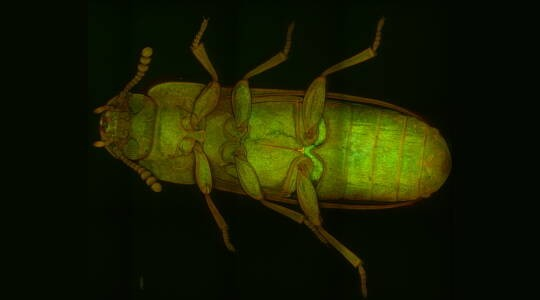
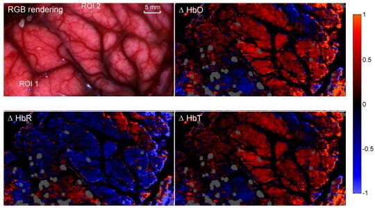
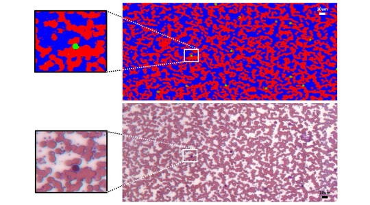
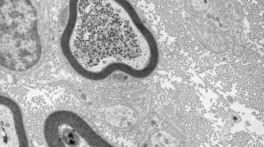
XIMEA cameras are suitable for both the conventional light optical microscopy applications and the fluorescence type of microscopy. They offer any required resolution while ensuring the highest image quality in real time, low noise and a high dynamic range. The cooled models allow long exposure times for low light conditions and reproducible results of examination for the finest of structures in a specimen that has to be analyzed.

Microscopy imaging systems application examples include biological and medical research, in life as well as in materials science fields.

XIMEA advantages

  • Highest resolutions

  • Increased Dynamic range

  • Strong sensitivity including NIR range

  • Longer exposure times

System integrators and manufacturers who used CCD based cameras in their devices are considering a switch to CMOS or sCMOS camera models due to the continuous discontinuation of CCD sensors with Sony CCD to be fully replaced by 2025.

Where CMOS is superior to CCD in speed and price, sCMOS is extraordinary in high sensitivity and low noise. Both are also eligible for integration into X-ray detector systems when coupled with fiber optic plate and scintillator coating.

They are also highly useful in case several sensors need to be combined in close proximity like for example on a single board.
This is what happened in the case of the Unity system from Aurox which carries out computer-assisted fluorescence microscopic examinations. 

The XIMEA team will be happy to talk directly about the specifics of your application to advise regarding the optimal camera choice.

Laser-free-confocal-fluorescence-microscopy
Laser free confocal - Spinning disk
The close proximity of dual sCMOS sensors placed on one board allowed the user to achieve a compact laser free confocal microscope system.
snapshot-hyperspectral-imaging-oxygenation-map-brain-surgery-in-vivo-fluorescence
In vivo hyperspectral fluorescence
The glow of fluorescent dyes is simulated via light from a particular wavelength and this light emission needs to be detected and captured.
hyperspectral-imaging-microscopy-pathology
Microscopy pathology
Fluorescence imaging coupled with hyperspectral qualities can distinguish even the smallest cellular and molecular structures, also for pathology.
Transmission-electron-scanning-microscopy-TEM-SEM-camera
TEM and SEM microscopy
Scanning and transmission electron microscopy are complex techniques that use an electron beam to form and capture the image of the specimen.

Cameras suitable for fluorescence microscopy applications

Want to discuss your fluorescence microscopy application? Get in touch!

We’re always curious to learn more about how you’re using XIMEA cameras and love discovering the most interesting applications to share with the world.

The XIMEA team would be very glad to collaborate with you on writing a case study and thus promote the experience among peers and highlight your case even further. Please fill out the form below to get started - we look forward to hearing from you!

contactdefault


Disclaimer

XIMEA is a component manufacturer - cameras being the main component and focus. 
We welcome your inquiries in regards to applications and will try to provide advice or recommendation to the best of our knowledge and experience.
However, XIMEA is not a Solution provider nor a System integrator, which means that we can offer suggestions only concerning the camera part of the overall setup.